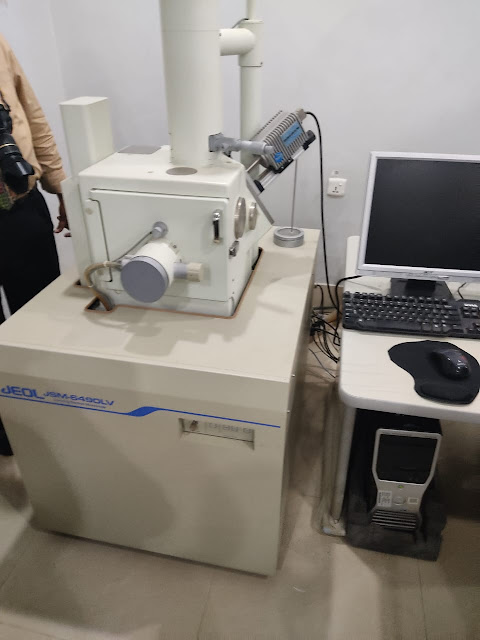
Electron microscopy (EM) is a powerful method employed to capture exceptionally detailed images of a wide range of specimens, both biological and non-biological. In the realm of biomedical research, EM serves as an invaluable tool for delving into the intricate architecture of tissues, cells, organelles, and macromolecular complexes.
The impressive level of resolution achieved with EM is attributed to its utilization of electrons, which possess incredibly short wavelengths, as the primary source of illumination. This capability opens a window into the finest details, often beyond the reach of conventional optical microscopy.
When it comes to exploring the microscopic world, electron microscopy collaborates with various supplementary techniques such as thin sectioning, immuno-labeling, and negative staining. This harmonious combination is driven by the pursuit of answers to specific scientific inquiries.
EM images offer profound insights into the structural foundations of cellular functionality and the underpinnings of various cellular diseases. It serves as an essential contributor to our comprehension of the microcosmic realm.
Within the realm of electron microscopy, two principal variants take center stage: the transmission electron microscope (TEM) and the scanning electron microscope (SEM). These instruments each have their unique strengths and applications in delivering critical visual information for the advancement of science.


0 Comments